Back Of Skull Anatomy : high quality 1:1 human skull model resin skeleton model ... - The muscles of the back that work together to support the spine, help keep the body upright and allow twist and bend in many directions.
Back Of Skull Anatomy : high quality 1:1 human skull model resin skeleton model ... - The muscles of the back that work together to support the spine, help keep the body upright and allow twist and bend in many directions.. The muscles of the back that work together to support the spine, help keep the body upright and allow twist and bend in many directions. Occipital bone anatomy the occipital bone is an unpaired bone, which covers the back of the head. As these bones grow throughout fetal and childhood development, they begin to fuse together, forming a single skull. Anatomy next provides anatomy learning tools for students and teachers The branching pattern of this artery forms readily visible grooves on the internal surface of the skull and these grooves can be traced back to their origin at the foramen spinosum.
Sep 22, 2020 · this human anatomy module is composed of diagrams, illustrations and 3d views of the back, cervical, thoracic and lumbar spinal areas as well as the various vertebrae. And it contributes to the action of chewing. The anatomy of the crown varies between different organisms. A large portion of the face is composed of the buccinator muscle, which compresses the cheek. Intermediate back muscles and c.
Smile! - Human skull on black background. | Human skull ... from i.pinimg.com As these bones grow throughout fetal and childhood development, they begin to fuse together, forming a single skull. Your back consists of three distinct layers of muscles, namely the superficial layer, the intermediate layer, and the deep layer. The physicians originally studying human anatomy thought the skull looked like an helmet. And it contributes to the action of chewing. The anatomy of the crown varies between different organisms. These layers of back muscles help to mobilize and stabilize your trunk during your day to day activities. The human crown is made of three layers of the scalp above the skull. The back muscles can be three types.
These layers of back muscles help to mobilize and stabilize your trunk during your day to day activities.
These layers of back muscles help to mobilize and stabilize your trunk during your day to day activities. The muscles of the back that work together to support the spine, help keep the body upright and allow twist and bend in many directions. The human crown is made of three layers of the scalp above the skull. Intermediate back muscles and c. Anatomy next provides anatomy learning tools for students and teachers It contains the osteology, arthrology and myology of the spine and back. Sep 22, 2020 · this human anatomy module is composed of diagrams, illustrations and 3d views of the back, cervical, thoracic and lumbar spinal areas as well as the various vertebrae. The back muscles can be three types. Occipital bone anatomy the occipital bone is an unpaired bone, which covers the back of the head. Deep back muscles superficial back muscles action movements of the shoulder. The anatomy of the crown varies between different organisms. A large portion of the face is composed of the buccinator muscle, which compresses the cheek. And it contributes to the action of chewing.
Occipital bone anatomy the occipital bone is an unpaired bone, which covers the back of the head. May 19, 2021 · anatomy of back muscles. Anatomy next provides anatomy learning tools for students and teachers The back muscles can be three types. As these bones grow throughout fetal and childhood development, they begin to fuse together, forming a single skull.
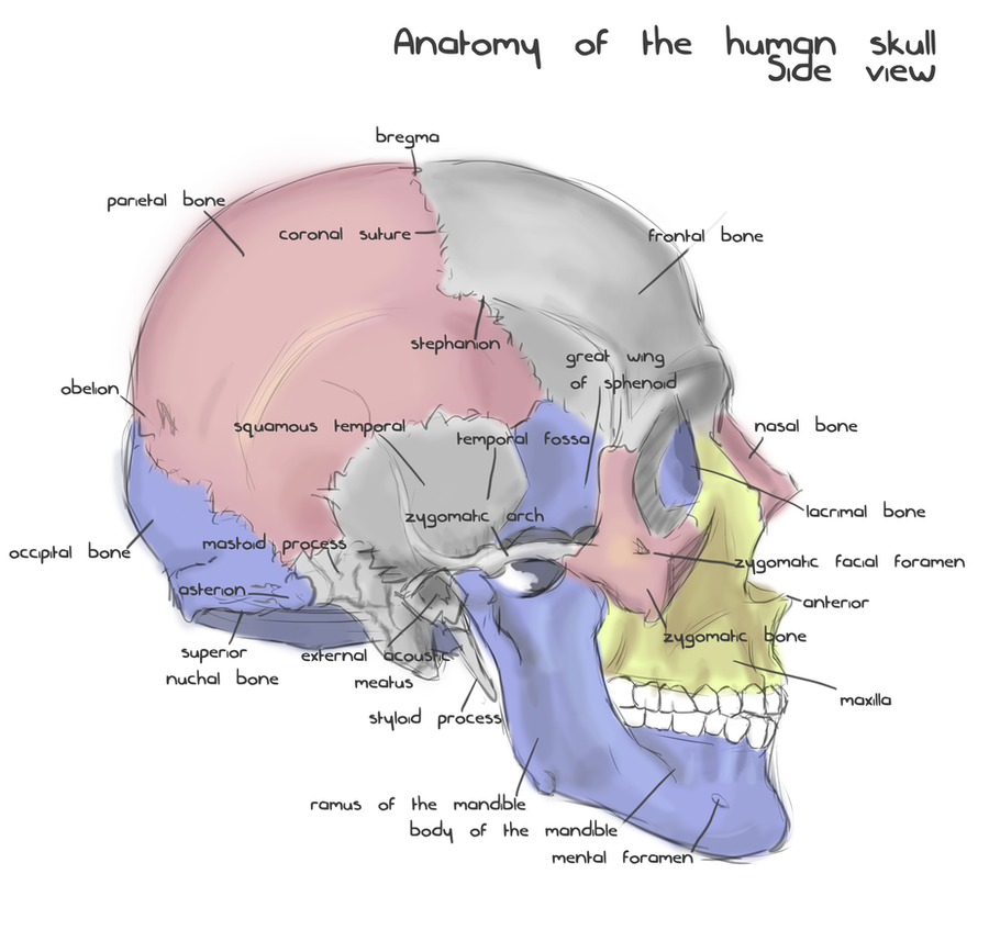
Annotated human skull anatomy - side view by shevans on ... from img00.deviantart.net It contains the osteology, arthrology and myology of the spine and back. The physicians originally studying human anatomy thought the skull looked like an helmet. During fetal development, the bones of the skull form within tough, fibrous membranes in a fetus' head. The back muscles can be three types. As these bones grow throughout fetal and childhood development, they begin to fuse together, forming a single skull. This muscle allows you to whistle, blow, and suck; The branching pattern of this artery forms readily visible grooves on the internal surface of the skull and these grooves can be traced back to their origin at the foramen spinosum. The anatomy of the crown varies between different organisms.
The back muscles can be three types.
It contains the osteology, arthrology and myology of the spine and back. Sep 22, 2020 · this human anatomy module is composed of diagrams, illustrations and 3d views of the back, cervical, thoracic and lumbar spinal areas as well as the various vertebrae. May 19, 2021 · anatomy of back muscles. Anatomy next provides anatomy learning tools for students and teachers Deep back muscles superficial back muscles action movements of the shoulder. Your back consists of three distinct layers of muscles, namely the superficial layer, the intermediate layer, and the deep layer. The crown also covers a range of bone sutures, and contains blood vessels and branches of the trigeminal nerve. The back muscles can be three types. The physicians originally studying human anatomy thought the skull looked like an helmet. The human crown is made of three layers of the scalp above the skull. This muscle allows you to whistle, blow, and suck; Intermediate back muscles and c. These layers of back muscles help to mobilize and stabilize your trunk during your day to day activities.
Deep back muscles superficial back muscles action movements of the shoulder. It contains the osteology, arthrology and myology of the spine and back. Anatomy next provides anatomy learning tools for students and teachers Occipital bone anatomy the occipital bone is an unpaired bone, which covers the back of the head. And it contributes to the action of chewing.
Most Popular in Skull Anatomy Page 256 from www.edoctoronline.com Sep 22, 2020 · this human anatomy module is composed of diagrams, illustrations and 3d views of the back, cervical, thoracic and lumbar spinal areas as well as the various vertebrae. A large portion of the face is composed of the buccinator muscle, which compresses the cheek. Anatomy next provides anatomy learning tools for students and teachers Intermediate back muscles and c. This muscle allows you to whistle, blow, and suck; The crown also covers a range of bone sutures, and contains blood vessels and branches of the trigeminal nerve. The physicians originally studying human anatomy thought the skull looked like an helmet. Occipital bone anatomy the occipital bone is an unpaired bone, which covers the back of the head.
During fetal development, the bones of the skull form within tough, fibrous membranes in a fetus' head.
The anatomy of the crown varies between different organisms. This muscle allows you to whistle, blow, and suck; Deep back muscles superficial back muscles action movements of the shoulder. The physicians originally studying human anatomy thought the skull looked like an helmet. The muscles of the back that work together to support the spine, help keep the body upright and allow twist and bend in many directions. As these bones grow throughout fetal and childhood development, they begin to fuse together, forming a single skull. Occipital bone anatomy the occipital bone is an unpaired bone, which covers the back of the head. The crown also covers a range of bone sutures, and contains blood vessels and branches of the trigeminal nerve. Intermediate back muscles and c. And it contributes to the action of chewing. Your back consists of three distinct layers of muscles, namely the superficial layer, the intermediate layer, and the deep layer. Sep 22, 2020 · this human anatomy module is composed of diagrams, illustrations and 3d views of the back, cervical, thoracic and lumbar spinal areas as well as the various vertebrae. Anatomy next provides anatomy learning tools for students and teachers


0 Komentar